ATSU takes the pulse of its anatomy programs
No bones about it, the anatomy labs at ATSU are pretty cool. From state-of-the-art computer software and a 3-D theatre to tried and true cadaver dissection and textbooks, the labs are a mighty source of knowledge and skill for students across disciplines. Both Arizona’s and Missouri’s facilities are finding new ways to integrate dissection and software, each putting student learning at the heart of its programs.
Anatomy labs get smart
Anatomy programs at ATSU are no strangers to change. Both programs have spent the last few years in transformation mode, questioning the status quo and asking tough questions about learning.
“Realizing that the goal of our institution is to create physicians in rural programs and family practice, the argument has been whether medical students who are not going into a surgical profession actually need cadaveric dissection,” explains Jay M. Crutchfield, MD, FACS, assistant professor and administrative anatomy director, SOMA.
“This begs for a change in the way we teach anatomy. Cadavers are still very useful, but they’re not the only way to teach.” — Dr. Crutchfield, SOMA
According to Dr. Crutchfield, a general surgeon trained in classical cadaveric dissection at Mount Sinai School of Medicine in New York, using cadavers to learn 3-D anatomy isn’t a necessity.
“When students become physicians they will really see anatomy by X-rays, CT scans, and MRIs and not necessarily how a surgeon would in the operating room,” he says. “This begs for a change in the way we teach anatomy. Cadavers are still very useful, but they’re not the only way to teach.”
This is why more technology was added to the program.
“An anatomy book is well-detailed, but it is not rotatable and you cannot piece apart the structures to appreciate the depths of the body,” says Joshua Menefee, OMS III. “Books and cadavers may not give us sufficient structures to learn from, and we will be using radiography to look at the body during our careers anyway.”

Students in Arizona experience anatomy as close to real life as possible with Cyber-Anatomy software in the 3-D theatre.
Second-year SOMA student Alicia Waite is grateful for the program’s emphasis on radiology.
“It has come in handy while reviewing for boards—especially anatomy via CT scans,” she says. “We are asked a ton of review questions about reading scans of different varieties.”
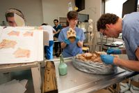
In Missouri, full-body cadaver dissection is emphasized. The lab, which was renovated little more than a year ago, is one of few in the country to operate solely on a body donation program.
“The course is heavily hands-on and dissection-based,” says KCOM Anatomy Chair Peter Kondrashov, PhD. “We probably have one of the best student-per-cadaver ratios in terms of number of students and the time students spend dissecting. We are probably in the top 5-10 percent [in the country].”
According to Dr. Kondrashov, dissection is a classical part of medical education and is an essential learning tool.
“No one has invented anything better than the human body to teach about the human body. I’ve looked into all kinds of things and still the human body is the most awesome teaching tool,” he says.
While dissection is pivotal to the program, technology plays a vital role. Dr. Kondrashov, who has been with KCOM since 2007 and chair since 2010, recognized that the lab was in need of a pick-me-up and that students could use a dose of engagement and relevance in class.
“We observed two worrying trends associated with the course for first-year medical students,” he explains. “The first was decline in anatomy sub-scores on the COMLEX 1 exam. The second was overall student dissatisfaction with the course.”
“We still rely heavily on what’s been the body and soul of the anatomy course for 100 years.” — Dr. Kondrashov, KCOM
To address these issues, the course was significantly restructured. Students were divided into two groups. One group dissects in the lab while the other receives a related clinical presentation by a local physician in the classroom or masters anatomical concepts using ultrasound. The groups then switch places so the whole class goes through the clinical case/ultrasound lab.
“The students said the introduction of clinical cases helped them learn the material better by putting it into clinical context,” Dr. Kondrashov says.
When possible, clinical cases incorporate medical imaging with related MRIs, X-rays, and CT scans to further illustrate anatomical structures. Portable ultrasounds were also added to the curriculum, allowing students to use this cutting-edge imaging technology on almost every part of the human body to learn “living” anatomy.
“The introduction of ultrasound was very helpful,” says Thomas Rountree, OMS II. “I saw ultrasound used several times during my preceptorship. Due to my training, I was able to follow along and recognize the images on screen.”
“Our efforts resulted in the implementation of a successful hybrid of a very traditional cadaver- based gross anatomy course with the best of available modern technology,” says Dr. Kondrashov. “We still rely heavily on what’s been the body and soul of the anatomy course for 100 years. We just take it a few steps further by adding technology and clinical correlations.”
Both campuses use online videos and several computer programs to aid student learning, such as Cyber-Anatomy, ToLTech VH Dissector, 4D Anatomy, and Online Dissector.

Arizona’s lab is used by many disciplines—medical, dental, physical therapy, athletic training, and physician assistant students all make use of the facility.
“As medical education questions the use of cadaveric dissection—and organ procurements and willed-body programs wane—technology is being used more and more,” says Dr. Crutchfield. “We use anatomical programs, true to human anatomy dissection, and actual operative videos, CT scans, ultrasounds, and radiographs from my own collection.”
Arizona’s recently updated computer system with an overhead high-definition camera and two projection screens means students can throw on a pair of 3-D glasses and get up close and personal with human anatomy without a scalpel.
“3-D technology allows us to show difficult anatomy that isn’t well seen during dissection,” explains Dr. Crutchfield.
The inner ear, the nervous system, and some of the musculoskeletal system are all explored using computer software.
Plastinates are another mainstay in Arizona’s lab. These teaching tools are odorless, don’t decay, and retain most of their original properties. The models are mostly used for teaching neuroanatomy in Missouri.
“I loved plastinates for learning intricate anatomy,” Menefee says. “They were key for observing some structures.”
Sue Hillman, MS, MA, ATC, who has spent more than 16 years as human anatomy director on the Arizona campus, says the greatest student learning tool right now is instruction via iTunes because it can be individualized and accessed any time.
“We strip out salient parts of surgery videos and provide those to our students on iTunes,” says Hillman. “Students who wish to review a video may download it and use it as they desire.”
Cadaveric dissection still plays a role in Arizona. In an optional dissection course, students who desire to learn by this method receive hands-on training. This group of 10-12 students meets once a week and prepares dissected specimens to be used in lectures with the rest of the class. They even occasionally assist faculty in instructing classmates.
ASDOH’s anatomy program, led by Wayne W. Cottam, DMD, MS, associate dean for community partnerships, also offers a dissection opportunity. Dental students take turns dissecting and teaching the dissection to classmates. According to Dr. Cottam, this allows students to learn the material quickly and integrate it to a level that they can teach it.
He feels that the combination of dissection and technology is integral to his class.
“You can learn the material you need from a book or software, and you can learn to do surgery without a dissecting a single thing,” Dr. Cottam says. “What a cadaver provides you is a chance to work.”

KCOM students take a closer look in the neuro anatomy lab.
Showing a backbone
Due to cuts in time and money, many programs across the country are shifting the way anatomy is taught.
“The traditional anatomy course is frequently being replaced with an integrated approach to medical education,” says SOMA Assistant Professor of Anatomy William Robinson, DPT, PhD. “An example of this is the clinical presentation curriculum that we use at SOMA.”
ATSU maintains that an intelligent fusion of software and dissection is the backbone to any successful program. After all, “You can’t take a cadaver home with you, but you can study Cyber- Anatomy on your home computer,” Dr. Robinson jests.
Both Missouri’s and Arizona’s anatomy programs highlight the need for students to learn in multifaceted ways as each uniquely combines traditional and up-and-coming methods to get the job done. Collaboration across campuses has been critical to each program’s success. Drs. Crutchfield and Kondrashov regularly share ideas and resources and are always on the lookout for ways to improve their programs.
Anatomy at ATSU is getting brainier—its labs unrestricted to one narrowminded track and both programs embracing new and old, history and future, and above all, connecting with the hearts and minds of students.